Shrinking the spotlight: super-resolution microscopy without labels
ARCNL researchers in the group of Peter Kraus have demonstrated a way to overcome the diffraction limit in optical microscopy, using laser techniques that shape light in both space and time. Published in the journal Optica, their method eliminates the need for fluorescent dyes or markers, making it a potential tool for applications from semiconductor manufacturing to clinical research and diagnostics.
Limits to light microscopy
Optical microscopes have transformed science and technology, but they come with a built-in limit: even the best lenses can only focus light into spots of a certain size. This so-called diffraction limit sets a hard boundary on how small visible details can be, or conversely how sharp an image can be.
In a new study published in Optica, researchers at ARCNL demonstrate a way to overcome this limit in a label-free microscopy method, meaning it does not rely on fluorescent dyes or markers. Instead, the team uses carefully shaped laser pulses to make the effective imaging spot much smaller, revealing details that would normally remain hidden.
Turning red light into blue light
The team’s method builds on a phenomenon called third-harmonic generation. Essentially, they shine extremely short laser pulses onto a sample, and the material responds by creating new light at a different color. For example, it turns red light into blue.
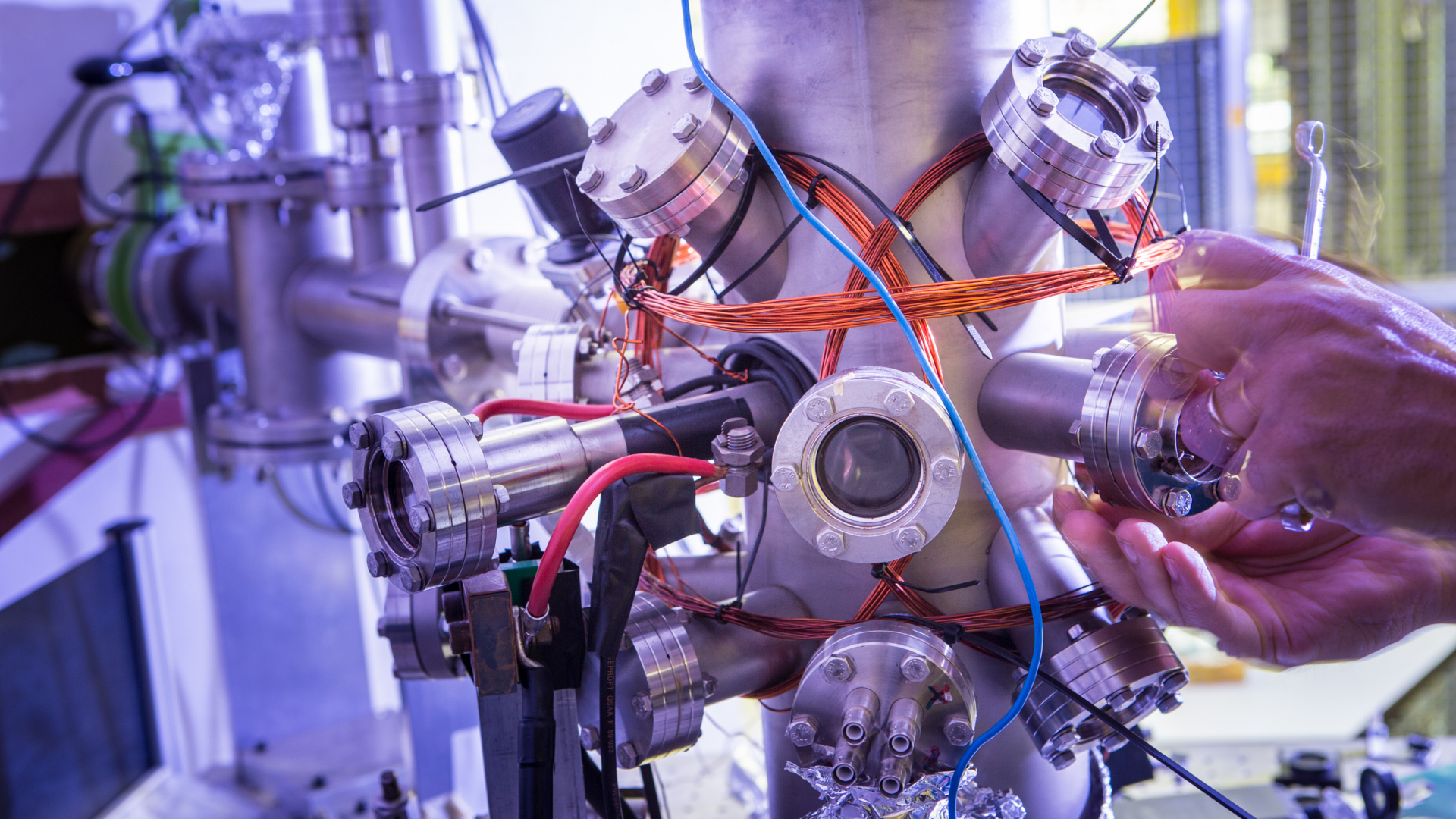
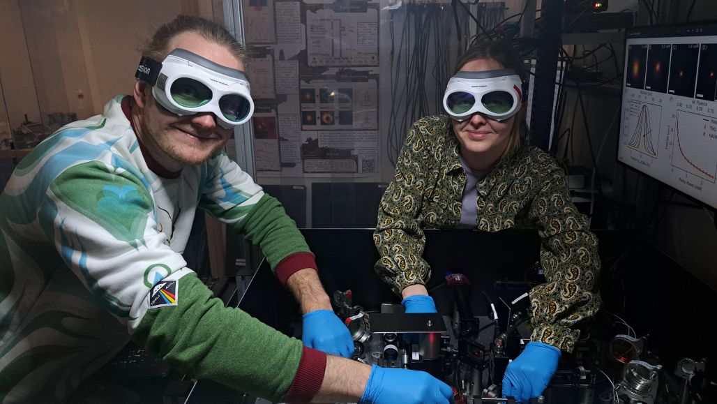
This type of color change is already useful in microscopy, but it normally still is bound by the diffraction limit. ARCNL researchers Kevin Murzyn, Tanya van Horen and Peter Kraus have found a way to push beyond that limit, by adding a second laser pulse that “switches off” the signal everywhere except in a tiny region.
A donut-shaped beam for sharper images
The key idea is to use a second laser beam with a donut-shaped profile. This beam suppresses the third-harmonic signal around the outside of the spot, leaving only a much smaller central region that contributes to the image.
With this approach, called HADES (Harmonic Deactivation Microscopy), the researchers show that the third-harmonic generation spot can be reduced to up to four times smaller than the diffraction limit.
Super-resolution for solid materials and clinical diagnostics
Many super-resolution microscopy techniques depend on fluorescent labels. While powerful, those labels are not always compatible with the types of samples and processes studied in physics and semiconductor research – the smallest features on semiconductor chips are much smaller than fluorescent labels.
The new approach offers a route to super-resolution imaging without these kinds of labels, which could be particularly valuable for a range of applications. In semiconductor inspection and metrology, adding labels is often not possible. Meanwhile, researchers in solid-state and condensed-matter physics want to observe fine structures in materials directly. Even in clinical settings, third-harmonic generation is already used for diagnosis and research, so being able to do super-resolution microscopy with this technique could improve diagnostic capabilities.
Kevin: “Seeing that we can break a fundamental optical limit without relying on labels, and that the theory we developed here fits nicely to the experiment, is the most exciting part of this project.”
By shaping light to have a smaller beam spot, this method of using HADES for super-resolution microscopy could support advancements in fields from solid-state physics and semiconductor manufacturing to clinical diagnostics. The team also points to the potential for imaging on extremely short timescales. Because the method uses ultrafast laser pulses in an extremely precise experimental setup, it could help enable studies of ultrafast dynamics – potentially even reaching attosecond timescales. This has possible benefits to developments in information transfer and energy storage.
Contact
For more information about this research, contact Peter Kraus
Reference
Kevin Murzyn, Tanya W. P. van Horen, & Peter M. Kraus, Control and scaling of nonlinear emission for super-resolution microscopy, Optica 13, 164-171 (2026).


