Visualizing the invisible: scientists push phase imaging beyond its limits
Researchers have developed a new imaging method that allows high-speed, label-free, and high-resolution phase imaging, using only a multimode optical fiber and a position-sensitive detector. This compact, camera-free technique surpasses traditional resolution limits and opens new doors for applications in biomedicine, semiconductor inspection, and beyond.
Reinventing phase imaging
Scientists at ARCNL, Vrije Universiteit Amsterdam, and CWI have developed a new technique that simplifies and accelerates quantitative phase imaging (QPI). Published in APL Photonics, their method eliminates the need for bulky, interferometric setups, offering high-resolution, label-free imaging through a compact fiber-based system.
What is phase imaging?
Most microscopes show how much light a sample absorbs, but many important samples – like living cells – are nearly transparent. Phase imaging reveals them by detecting tiny changes in how light bends or slows down as it passes through. This makes invisible structures visible, without needing dyes or labels.
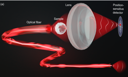
The new approach uses a standard multimode optical fiber (MMF), which naturally produces complex random speckle illumination. Combined with a position-sensitive detector and a simple lens, this setup enables high-resolution reconstruction of both the phase and amplitude of a sample — without reference beams, or iterative post-processing.
Why this matters
This development opens the door to a new class of imaging tools that are fast, precise, and remarkably compact. It holds particular promise for:
- Biomedical research, enabling non-invasive, high-speed imaging of living cells
- Semiconductor manufacturing, where nanoscale defects must be identified with extreme precision
- Fiber-optic endoscopy, allowing deep-tissue imaging with minimal invasiveness
The system achieves video-rate performance up to 50 frames per second, using only a scanning mirror and a pre-calibrated detector. No camera is required during imaging, which simplifies both the setup and data acquisition. Ultimately, the imaging speed is limited only by the scanning rate of the illumination device, not by the computational or detection steps.
Looking ahead
With further development, the technique could be integrated into next-generation microscopes, endoscopes, or industrial inspection tools. Its simplicity makes it a strong candidate for embedded imaging chips or automated diagnostics. The researchers also envision combining their method with advanced computational techniques or machine learning to enhance resolution even further — making this a future-proof step toward faster, smarter, and more accessible imaging technologies.
Reference
Aleksandra Ivanina, Maxim Marshall, Ksenia Abrashitova, Tristan van Leeuwen and Lyubov V. Amitonova, Quantitative phase imaging with a multimode fiber, APL Photonics, April 21 (2025).
Find out more in the research paper: Quantitative phase imaging with a multimode fiber


