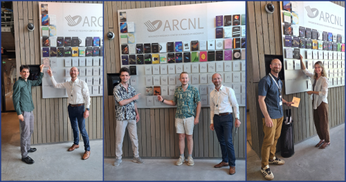
Four new Doctors add their covers to the Theses Wall this summer
Paul Ducarme
On May 4, joint AMOLF/ARCNL PhD candidate Paul Ducarme successfully defended his thesis, “Harnessing Mechanical Instabilities for Functional Structures Using Nonlinear Building Blocks.” For his doctoral research, Paul developed elastic structures to demonstrate new phenomena in mechanics using instabilities. He says: “I studied how shape and geometry can be tuned to create new functional behaviors in matter, using only mechanics. I constructed the systems with simple building blocks, like a LEGO set.”
Maksym Illienko
On June 8, Maksym Illienko successfully defended his PhD thesis. Supervised by Professor Stefan Witte, his thesis is titled “Picosecond ultrasonics for nanoscale subsurface structural characterization.” In it, he details how sound can be made from light and used to detect hidden structures at the nano scale. Maksym’s research involved two parts: generating sound waves from light, also known as photoacoustics, and nanoscale imaging of structures hidden behind opaque layers. He was able to show that, under certain conditions, these nanoscale structures could be imaged using sound.
Aleksandra Ivanina
On June 23, Aleksandra Ivanina successfully defended her PhD thesis! She’s proudly added the cover of her thesis to the ARCNL Theses Wall. Her thesis is titled, ” Engineering Illumination Through Multimode Fibers for Imaging and Metrology.” Supervised by Dr. Lyuba Amitonova, Aleksandra’s doctoral research focused on controlling light using optical fibers. Looking at a variety of practical applications, her research showed how powerful and versatile optical fibers are as tools for working with light.
Kevin Murzyn
On July 1, Kevin Murzyn successfully defended his PhD thesis and added his thesis cover to the ARCNL Theses Wall. Supervised by group leader Dr. Peter Kraus, Kevin’s doctoral research focused on super-resolution microscopy using high-harmonic generation. His technique, Harmonic Deactivation Microscopy (HADES), uses carefully shaped laser pulses to make the effective imaging spot much smaller, revealing details that would normally remain hidden.
The method overcomes the so-called diffraction limit for optical microscopy without relying on fluorescent dyes or markers. “In principle, it can be scaled down to nanometer-level resolution,” says Kevin, “but we weren’t able to show that yet. There’s more work to be done to make that happen.”